Glowscope
Context:
- In 2014, a group of scientists at the Stanford University released Foldscope, a handheld microscope made almost entirely out of paper, which takes 30 minutes to put together, and which could capture images of cells.
- Costs less than 400, So far, millions of people, especially schoolchildren around the world have taken images of the microscopic world with Foldscopes, while dozens of scientific studies have been conducted with the help of this instrument.
- Foldscope democratised the world’s access to optical microscopy. Now, researchers at the Winona State University, Minnesota, have created a design for a ‘glowscope’, a device that could democratise access to fluorescence microscopy — at least partly so.
What is fluorescence microscopy?
- An optical microscope views an object by studying how it absorbs, reflects or scatters visible light.
- A fluorescence microscope views an object by studying how it re-emits light that it has absorbed, i.e. how it fluoresces. This is its basic principle.
- The object is illuminated with light of a specific wavelength. Particles in the object absorb this light and re-emit it at a higher wavelength (i.e. different colour). These particles are called fluorophores; the object is infused with them before being placed under the microscope.
- There are versions of fluorescent microscopes with more sophisticated abilities, such as epifluorescence and confocal laser-scanning microscopes.
- When the fluorophores fluoresce, a fluorescent microscope can track them as they move inside the object, revealing the object’s internal shape and other characteristics.
- For instance, a fluorophore called the Hoechst stain binds to the DNA and is excited by ultraviolet light.
- So, a tissue sample collected from a person could be injected with the Hoechst stain and placed under a fluorescent microscope. When the sample is illuminated by ultraviolet light, the stain absorbs the light and re-emits it at a higher wavelength.
- The microscope will point out where this is happening: in the nuclei of cells, where DNA is located. This way, the nuclei in the tissue can be labelled for further study.
- Scientists have developed different fluorophores to identify and study different entities, from specific parts of the DNA to protein complexes.
- On the negative side, fluorescence microscopes cost at least a lakh rupee, but often up to crores.
How does the new device improve access?
- In the new study, researchers from the Winona State University have described a rudimentary fluorescence microscope that they say can be put together at a cost of $30-$50 (₹2,500-₹4,100).
- The scientists demonstrate the ability of these devices to detect green and red fluorophores and to monitor and detect changes to heart rate and rhythmicity in embryonic zebrafish.”
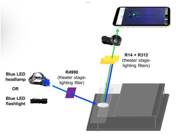
- Their set-up consists of two plexiglass surfaces, an LED flashlight, three theatre stage-lighting filters, a clip-on macro lens, and a smartphone.
- The smartphone (with the lens attached) is placed on one surface that is suspended at a height (say, a foot above). The second sheet is placed below and holds the object.
- In their study, the objects were zebrafish embryos in a petri dish, prepared according to well-established guidelines to ensure they aren’t harmed.
- They were injected with different fluorophores depending on which part of the embryos were of interest. The sources of illumination were also LED flashlights emitting light of correspondingly different wavelengths.
- One of the stage-lighting filters was held between the flashlight and the object and the other two were held between the object and the smartphone.
- The role of these filters was to ensure that light of the right frequency reached the object and that fluoresced light of a suitable frequency reached the camera.
- With this setup, the researchers were able to image the creatures’ brain, spinal cord (using a fluorophore called DsRed), heart (mCherry), and head and jaw bones (mRFP).
- They were able to zoom in and out using the smartphone camera and the clip-on lens, and by adjusting the distance between the sample and the smartphone platforms.
| Practice Question
1. What is fluorescence microscopy? What role Glowscope can play in making it reachable to all? |